HLA-DRB4: A Promising Drug Target / Biomarker (G3126)


HLA-DRB4: A Promising Drug Target / Biomarker
HLA-DRB4 is a human leukocyte antigen (HLA) gene that is located on chromosome 6p21. It is a key regulator of immune responses and has been implicated in a number of autoimmune diseases, including rheumatoid arthritis, lupus, and multiple sclerosis. In addition, HLA-DRB4 has also been identified as a potential drug target and biomarker for a number of diseases.
The HLA-DRB4 gene is located on chromosome 6p21 and is responsible for the production of theHLA-DRB4 protein. This protein plays a critical role in the immune response by helping to recognize and respond to foreign substances in the body.
Implications for Autoimmune Diseases
HLA-DRB4 is a key regulator of the immune response and has been implicated in a number of autoimmune diseases. One of the most well-known examples is rheumatoid arthritis (RA), which is an autoimmune disease that causes joint inflammation and pain.
Research has shown that HLA-DRB4 is expressed in the cells of individuals with RA and that it is involved in the development of the disease. Additionally, studies have shown that individuals with RA have lower levels of HLA-DRB4 in their blood than healthy individuals.
Another example of an autoimmune disease that is associated with HLA-DRB4 is lupus, a chronic autoimmune disease that can affect multiple organs and systems in the body.
Research has shown that HLA-DRB4 is expressed in the cells of individuals with lupus and that it is involved in the development of the disease. Additionally, studies have shown that individuals with lupus have lower levels of HLA-DRB4 in their blood than healthy individuals.
HLA-DRB4 as a Potential Drug Target
HLA-DRB4 has also been identified as a potential drug target for a number of diseases. One of the most promising targets is cancer, as HLA-DRB4 has been shown to be involved in the regulation of cancer cell growth and survival.
Research has shown that HLA-DRB4 is expressed in a variety of cancer cells and that it is involved in the development and progression of cancer. Additionally, studies have shown that inhibiting HLA-DRB4 can lead to a reduction in cancer cell growth and survival.
Another potential drug target for HLA-DRB4 is neurodegenerative diseases, such as Alzheimer's disease.
Research has shown that HLA-DRB4 is expressed in the cells of individuals with Alzheimer's disease and that it is involved in the development and progression of the disease. Additionally, studies have shown that inhibiting HLA-DRB4 can lead to a reduction in the production of beta-amyloid plaques, a hallmark of Alzheimer's disease.
HLA-DRB4 as a Biomarker
HLA-DRB4 has also been identified as a potential biomarker for a number of diseases. One of the most promising applications for HLA-DRB4 as a biomarker is its potential use as a diagnostic marker for multiple sclerosis (MS).
Research has shown that HLA-DRB4 is expressed in the cells of individuals with MS and that it is involved in the development and progression of the disease. Additionally, studies have shown that individuals with MS have lower levels of HLA-DRB4 in their blood than healthy individuals.
HLA-DRB4 has also been used as a biomarker for other autoimmune diseases, including rheumatoid arthritis and lupus.
Conclusion
HLA-DRB4 is a human leukocyte antigen that is located on chromosome 6p21 and plays a critical role in the immune response. It is also
Protein Name: Major Histocompatibility Complex, Class II, DR Beta 4
Functions: Binds peptides derived from antigens that access the endocytic route of antigen presenting cells (APC) and presents them on the cell surface for recognition by the CD4 T-cells. The peptide binding cleft accommodates peptides of 10-30 residues. The peptides presented by MHC class II molecules are generated mostly by degradation of proteins that access the endocytic route, where they are processed by lysosomal proteases and other hydrolases. Exogenous antigens that have been endocytosed by the APC are thus readily available for presentation via MHC II molecules, and for this reason this antigen presentation pathway is usually referred to as exogenous. As membrane proteins on their way to degradation in lysosomes as part of their normal turn-over are also contained in the endosomal/lysosomal compartments, exogenous antigens must compete with those derived from endogenous components. Autophagy is also a source of endogenous peptides, autophagosomes constitutively fuse with MHC class II loading compartments. In addition to APCs, other cells of the gastrointestinal tract, such as epithelial cells, express MHC class II molecules and CD74 and act as APCs, which is an unusual trait of the GI tract. To produce a MHC class II molecule that presents an antigen, three MHC class II molecules (heterodimers of an alpha and a beta chain) associate with a CD74 trimer in the ER to form a heterononamer. Soon after the entry of this complex into the endosomal/lysosomal system where antigen processing occurs, CD74 undergoes a sequential degradation by various proteases, including CTSS and CTSL, leaving a small fragment termed CLIP (class-II-associated invariant chain peptide). The removal of CLIP is facilitated by HLA-DM via direct binding to the alpha-beta-CLIP complex so that CLIP is released. HLA-DM stabilizes MHC class II molecules until primary high affinity antigenic peptides are bound. The MHC II molecule bound to a peptide is then transported to the cell membrane surface. In B-cells, the interaction between HLA-DM and MHC class II molecules is regulated by HLA-DO. Primary dendritic cells (DCs) also to express HLA-DO. Lysosomal microenvironment has been implicated in the regulation of antigen loading into MHC II molecules, increased acidification produces increased proteolysis and efficient peptide loading
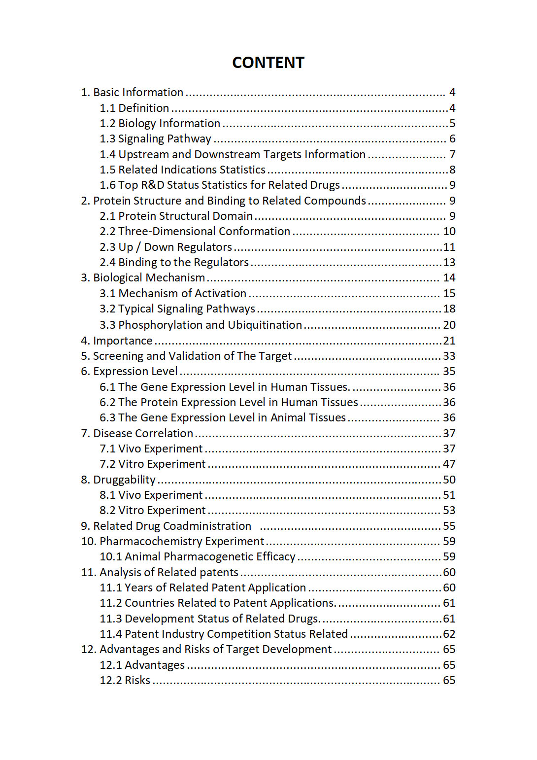
The "HLA-DRB4 Target / Biomarker Review Report" is a customizable review of hundreds up to thousends of related scientific research literature by AI technology, covering specific information about HLA-DRB4 comprehensively, including but not limited to:
• general information;
• protein structure and compound binding;
• protein biological mechanisms;
• its importance;
• the target screening and validation;
• expression level;
• disease relevance;
• drug resistance;
• related combination drugs;
• pharmacochemistry experiments;
• related patent analysis;
• advantages and risks of development, etc.
The report is helpful for project application, drug molecule design, research progress updates, publication of research papers, patent applications, etc. If you are interested to get a full version of this report, please feel free to contact us at BD@silexon.ai
More Common Targets
HLA-DRB5 | HLA-DRB6 | HLA-DRB7 | HLA-DRB8 | HLA-DRB9 | HLA-E | HLA-F | HLA-F-AS1 | HLA-G | HLA-H | HLA-J | HLA-K | HLA-L | HLA-N | HLA-P | HLA-U | HLA-V | HLA-W | HLCS | HLF | HLTF | HLX | HM13 | HMBOX1 | HMBS | HMCES | HMCN1 | HMCN2 | HMG20A | HMG20B | HMGA1 | HMGA1P2 | HMGA1P4 | HMGA1P7 | HMGA1P8 | HMGA2 | HMGA2-AS1 | HMGB1 | HMGB1P1 | HMGB1P10 | HMGB1P19 | HMGB1P37 | HMGB1P38 | HMGB1P46 | HMGB1P5 | HMGB1P6 | HMGB2 | HMGB2P1 | HMGB3 | HMGB3P1 | HMGB3P14 | HMGB3P15 | HMGB3P19 | HMGB3P2 | HMGB3P22 | HMGB3P24 | HMGB3P27 | HMGB3P30 | HMGB3P6 | HMGB4 | HMGCL | HMGCLL1 | HMGCR | HMGCS1 | HMGCS2 | HMGN1 | HMGN1P16 | HMGN1P30 | HMGN1P37 | HMGN1P8 | HMGN2 | HMGN2P13 | HMGN2P15 | HMGN2P18 | HMGN2P19 | HMGN2P24 | HMGN2P25 | HMGN2P30 | HMGN2P38 | HMGN2P46 | HMGN2P5 | HMGN2P6 | HMGN2P7 | HMGN3 | HMGN3-AS1 | HMGN4 | HMGN5 | HMGXB3 | HMGXB4 | HMHB1 | HMMR | HMOX1 | HMOX2 | HMSD | HMX1 | HMX2 | HNF1A | HNF1A-AS1 | HNF1B | HNF4A


