PDIA3: A promising drug target and biomarker for the treatment of various diseases


PDIA3: A promising drug target and biomarker for the treatment of various diseases
The endoplasmic reticulum (ER) is a complex organelle that plays a crucial role in the regulation of protein synthesis, quality control, and degradation. One of the key proteins involved in this process is PDIA3, which is a non-coding RNA molecule that is expressed in high levels in the ER and is known to play a critical role in the regulation of protein translation. PDIA3 has also been implicated in the development and progression of various diseases, making it an attractive drug target and biomarker. In this article, we will explore the biology of PDIA3, its potential as a drug target, and its potential as a biomarker for the diagnosis and treatment of various diseases.
PDIA3: Structure and function
PDIA3 is a 14 kDa protein that is synthesized primarily in the ER. It is composed of a unique alternating double helix structure that consists of a 尾-sheet and a 尾-halo region. The 尾-sheet is responsible for the protein's stability and functions as a binding site, while the 尾-halo region interacts with other proteins and molecules in the ER.
PDIA3 is involved in the regulation of protein translation by interacting with several key protein molecules, including the 40S ribosome complex, the transfer RNA, and the elongation factor. It has been shown that PDIA3 can interact with the 40S ribosome complex and the transfer RNA, allowing it to regulate the loading of new proteins onto the ribosome and the selection of specific target genes for translation.
PDIA3 is also involved in the regulation of protein degradation by interacting with the protein degradation machinery. It has been shown that PDIA3 can interact with the 26S proteasome, the key protein involved in the degradation of protein via the proteasome pathway. This interaction between PDIA3 and the proteasome suggests that PDIA3 may play a role in the regulation of protein degradation and may be a potential drug target for diseases that are characterized by the accumulation of abnormally modified or misfolded proteins.
PDIA3's role in disease
The accumulation of misfolded or abnormally modified proteins is a hallmark of various diseases, including neurodegenerative disorders, cancer, and diseases associated with protein misfolding, such as amyloidosis. PDIA3 has been shown to be involved in the regulation of the misfolding of proteins and the accumulation of misfolded proteins in these diseases.
For example, PDIA3 has been shown to play a role in the regulation of the misfolding of the protein huntingtin (HING) in neurodegenerative disorders. HING is a protein that has been implicated in the development of various neurodegenerative disorders, including Alzheimer's disease and Parkinson's disease. Studies have shown that PDIA3 can interact with HING and regulate its misfolding, suggesting that it may play a role in the development and progression of these diseases.
PDIA3 has also been shown to be involved in the regulation of the misfolding of the protein wnt-1 in cancer. Wnt-1 is a protein that has been implicated in the development and progression of various cancers, including breast and colon cancer. Studies have shown that PDIA3 can interact with Wnt-1 and regulate its misfolding, suggesting that it may play a role in the development and progression of these cancers.
PDIA3 as a drug target
PDIA3's involvement in the regulation of protein translation and degradation makes it an attractive drug target for the treatment of various diseases. Several studies have shown that inhibiting PDIA3 can be effective in treating diseases associated with the accumulation of misfolded or abnormally modified proteins.
For example, studies have shown that inhibiting PDIA3 can
Protein Name: Protein Disulfide Isomerase Family A Member 3
Functions: Disulfide isomerase which catalyzes the formation, isomerization, and reduction or oxidation of disulfide bonds (PubMed:7487104, PubMed:27897272). Associates with calcitriol, the active form of vitamin D3 which mediates the action of this vitamin on cells (PubMed:27897272). Association with calcitriol does not affect its enzymatic activity (PubMed:27897272)
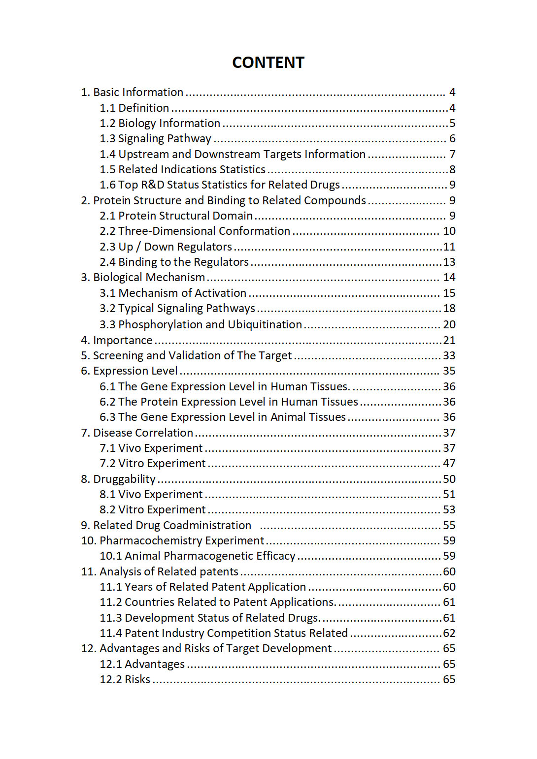
The "PDIA3 Target / Biomarker Review Report" is a customizable review of hundreds up to thousends of related scientific research literature by AI technology, covering specific information about PDIA3 comprehensively, including but not limited to:
• general information;
• protein structure and compound binding;
• protein biological mechanisms;
• its importance;
• the target screening and validation;
• expression level;
• disease relevance;
• drug resistance;
• related combination drugs;
• pharmacochemistry experiments;
• related patent analysis;
• advantages and risks of development, etc.
The report is helpful for project application, drug molecule design, research progress updates, publication of research papers, patent applications, etc. If you are interested to get a full version of this report, please feel free to contact us at BD@silexon.ai
More Common Targets
PDIA3P1 | PDIA4 | PDIA5 | PDIA6 | PDIK1L | PDILT | PDK1 | PDK2 | PDK3 | PDK4 | PDLIM1 | PDLIM1P4 | PDLIM2 | PDLIM3 | PDLIM4 | PDLIM5 | PDLIM7 | PDP1 | PDP2 | PDPK1 | PDPK2P | PDPN | PDPR | PDPR2P | PDRG1 | PDS5A | PDS5B | PDS5B-DT | PDSS1 | PDSS2 | PDX1 | PDXDC1 | PDXDC2P-NPIPB14P | PDXK | PDXP | PDYN | PDYN-AS1 | PDZD11 | PDZD2 | PDZD4 | PDZD7 | PDZD8 | PDZD9 | PDZK1 | PDZK1IP1 | PDZK1P1 | PDZPH1P | PDZRN3 | PDZRN3-AS1 | PDZRN4 | PEA15 | PEAK1 | PEAK3 | PEAR1 | PeBoW complex | PEBP1 | PEBP1P2 | PEBP4 | PECAM1 | PECR | PEDS1 | PEDS1-UBE2V1 | PEF1 | PEG10 | PEG13 | PEG3 | PEG3-AS1 | PELATON | PELI1 | PELI2 | PELI3 | PELO | PELP1 | PELP1-DT | PEMT | PENK | PENK-AS1 | PEPD | Peptidyl arginine deiminase (PAD) | Peptidylprolyl Isomerase | PER1 | PER2 | PER3 | PER3P1 | PERM1 | Peroxiredoxin | Peroxisome Proliferator-Activated Receptors (PPAR) | PERP | PES1 | PET100 | PET117 | PEX1 | PEX10 | PEX11A | PEX11B | PEX11G | PEX12 | PEX13 | PEX14 | PEX16


