Drug Target and Biomarker: MCL-1 (Mcl-1) plays a crucial role in cell survival and apoptosis regulation


Drug Target and Biomarker: MCL-1 (Mcl-1) plays a crucial role in cell survival and apoptosis regulation
ERK inhibition leads to GSK3beta-mediated Mcl-1 phosphorylation, promoting apoptosis through BH3 liberation and/or FBW7-mediated degradation.
Phosphorylation of Mcl-1 at Ser13 partially masks its BH3 domain, inhibiting the interaction with Noxa and potentially preventing apoptosis.
MCL-1 binds to and prevents the phosphorylation of Cofilin, allowing cytoskeletal invasion by cancer cells. The inhibition of MCL-1, along with SRC family kinase inhibitors, can suppress invasion, induce cell death, and increase drug sensitivity.
In MYCN-amplified SCLC, N-Myc inhibits Bim. Treatment with JQ1 decreases N-Myc levels, leading to the up-regulation of Bim. Combining JQ1 with ABT-263 disrupts the interaction between Bim and BCL-2/Mcl-1, promoting Bim release and apoptosis.
Metformin treatment affects the interactions involving Mcl-1 in DLD-1 cells. It increases the interaction between Noxa and Mcl-1, decreases the interactions between Mcl-1 and Mule, and inhibits Mcl-1 polyubiquitination.
These findings highlight the complex regulation of Mcl-1 in various cellular processes, including apoptosis, invasion, and drug sensitivity. The understanding of the mechanisms involved in Mcl-1 regulation can potentially lead to the development of therapeutic strategies targeting Mcl-1 for cancer treatment.
Based on the provided context information, the following key viewpoints can be extracted:
BIM/MCL-1 ChNPs: The synthesis of BIM/MCL-1 ChNPs involves the encapsulation of BIM-encoding AAV and MCL-1 siRNA in an acid-degradable polyketal shell. The PK shell is designed to degrade in the mildly acidic environment of endosomes/lysosomes, releasing the siRNA and AAV for intracellular processes.
Synergistic effects of BIM expression and MCL-1 silencing: The BIM/MCL-1 ChNPs facilitate the simultaneous expression of BIM and silencing of MCL-1 in BCR-ABL+ leukemia cells. This synergistic combination leads to the re-sensitization of cells to BIM-mediated apoptosis and suppression of cell proliferation.
Flavivirus infection and MCL1 regulation: Flavivirus infection results in the reduction of MCL1 protein. Delayed apoptosis in normal cells, mediated by BCLXL, allows the virus to spread to neighboring cells, increasing pathogenicity. Inhibition of BCLXL accelerates apoptosis and prevents viral dissemination by removing infected cells through phagocytosis.
USP24 and Mcl-1 in T-ALL: USP24 plays a crucial role in the survival of T-ALL cells by deubiquitinating Mcl-1, which helps maintain mitochondrial integrity. WP1130, a compound that targets USP24, inhibits its deubiquitination activity, leading to increased degradation of Mcl-1. This ultimately induces apoptosis of T-ALL cells.
TRAF6, p53, and MCL-1 in genotoxic stress: Before genotoxic stress, TRAF6-mediated ubiquitination of p53 inhibits its binding to BAK/MCL-1, keeping p53 away from mitochondria. Upon genotoxic stress, p53 levels increase, TRAF6 levels decrease, and p53 translocates to mitochondria for BAK oligomerization, apoptosis, and expression of p21 and GADD45 genes. TRAF6 deficiency leads to the accumulation of p53 in mitochondria, triggering apoptosis even in the absence of stress.
Combination therapy targeting BCL-2 family proteins: In untreated cells, pro-survival BCL-2, BCL-XL, and MCL-1 inhibit pro-apoptotic BAX and BAK proteins. Combination therapies, such as using NOXA or fenretinide with ABT-263, disrupt the interaction between BAK and MCL-1 and inhibit the function of BCL-XL, leading to enhanced cell death.
These viewpoints provide insights into the roles and regulation of MCL1 in various contexts, including nanoparticle-based drug delivery, viral infections, cancer therapy, and response to genotoxic stress.
Protein Name: MCL1 Apoptosis Regulator, BCL2 Family Member
Functions: Involved in the regulation of apoptosis versus cell survival, and in the maintenance of viability but not of proliferation. Mediates its effects by interactions with a number of other regulators of apoptosis. Isoform 1 inhibits apoptosis. Isoform 2 promotes apoptosis
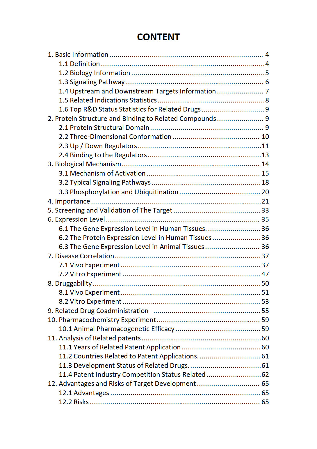
The "MCL1 Target / Biomarker Review Report" is a customizable review of hundreds up to thousends of related scientific research literature by AI technology, covering specific information about MCL1 comprehensively, including but not limited to:
• general information;
• protein structure and compound binding;
• protein biological mechanisms;
• its importance;
• the target screening and validation;
• expression level;
• disease relevance;
• drug resistance;
• related combination drugs;
• pharmacochemistry experiments;
• related patent analysis;
• advantages and risks of development, etc.
The report is helpful for project application, drug molecule design, research progress updates, publication of research papers, patent applications, etc. If you are interested to get a full version of this report, please feel free to contact us at BD@silexon.ai
More Common Targets
MCM10 | MCM2 | MCM3 | MCM3AP | MCM3AP-AS1 | MCM4 | MCM5 | MCM6 | MCM7 | MCM8 | MCM8-MCM9 complex | MCM9 | MCMBP | MCMDC2 | MCOLN1 | MCOLN2 | MCOLN3 | MCPH1 | MCPH1-AS1 | MCPH1-DT | MCRIP1 | MCRIP2 | MCRS1 | MCTP1 | MCTP2 | MCTS1 | MCTS2 | MCU | MCUB | MCUR1 | MDC1 | MDFI | MDFIC | MDGA1 | MDGA2 | MDH1 | MDH1B | MDH2 | MDK | MDM1 | MDM2 | MDM4 | MDN1 | MDS2 | ME1 | ME2 | ME3 | MEA1 | MEAF6 | MEAF6P1 | MEAK7 | Mechanoelectrical transducer (MET) channel | Mechanosensitive Ion Channel | MECOM | MECOM-AS1 | MeCP1 histone deacetylase (HDAC) complex | MECP2 | MECR | MED1 | MED10 | MED11 | MED12 | MED12L | MED13 | MED13L | MED14 | MED14P1 | MED15 | MED15P8 | MED16 | MED17 | MED18 | MED19 | MED20 | MED21 | MED22 | MED23 | MED24 | MED25 | MED26 | MED27 | MED28 | MED29 | MED30 | MED31 | MED4 | MED4-AS1 | MED6 | MED7 | MED8 | MED9 | MEDAG | Mediator Complex | Mediator of RNA Polymerase II Transcription | MEF2A | MEF2B | MEF2C | MEF2C-AS1 | MEF2C-AS2 | MEF2D


