Targeting TYR (OCAIA), a Drug Target and Biomarker for the Treatment of Neurodegenerative Disorders


Targeting TYR (OCAIA), a Drug Target and Biomarker for the Treatment of Neurodegenerative Disorders
Neurodegenerative disorders are a group of debilitating and often fatal brain diseases that affect millions of people worldwide. These disorders include Alzheimer's disease, Parkinson's disease, Huntington's disease, and other forms of dementia. The development of these disorders is closely associated with the accumulation of toxic substances, including protein aggregates, in the brain. One of the most promising strategies to treat neurodegenerative disorders is to target the pathways that are responsible for these accumulations. TYR (Ocaina) is a protein that has been identified as a potential drug target and biomarker for the treatment of neurodegenerative disorders. In this article, we will explore the biology of TYR and its potential as a drug target and biomarker.
The biology of TYR
TYR is a 21-kDa protein that is expressed in many tissues, including brain, heart, and liver. It is a member of the TYR family, which includes several related proteins that are involved in the regulation of cell signaling pathways. TYR is composed of a unique transmembrane domain, a catalytic domain, and an intracellular tail.
The transmembrane domain of TYR is characterized by a unique arrangement of amino acids that are involved in the formation of a ion channel. This channel allows TYR to regulate the movement of ions into and out of the cell, which is critical for maintaining the integrity of the cell membrane and the stability of the cell's signaling pathways. The catalytic domain of TYR contains a catalytic active site that is responsible for the chemical reaction that enables TYR to regulate cell signaling pathways.
In addition to its transmembrane and catalytic domains, TYR also has an intracellular tail that is involved in the regulation of its stability and localization to the cell surface. The intracellular tail of TYR is composed of a unique protein called TYR-TN, which contains a critical region that is involved in the formation of a protein-protein interaction network.
The potential drug target of TYR
The accumulation of toxic substances, including protein aggregates, in the brain is a major contributor to the development and progression of neurodegenerative disorders. TYR has been shown to play a role in the regulation of protein aggregation and may be a potential drug target for the treatment of neurodegenerative disorders.
One of the mechanisms by which TYR may contribute to the regulation of protein aggregation is by modulating the formation of protein-protein interfaces. Protein-protein interfaces are critical for the regulation of protein structure and function, and are often involved in the formation of protein aggregates. TYR has been shown to play a role in the regulation of protein-protein interfaces by modulating the formation of a protein-protein interaction network in the intracellular tail of TYR.
In addition to its potential role in modulating protein-protein interfaces, TYR has also been shown to play a role in the regulation of protein aggregation by modulating the stability of TYR itself. TYR is a protein that is involved in the regulation of ion channels in the cell membrane, and it has been shown to play a role in the regulation of the stability of these channels. The regulation of ion channels is closely associated with the regulation of protein aggregation, and TYR may be involved in the regulation of protein aggregation by modulating the stability of its own structure.
The potential biomarker status of TYR
The potential use of TYR as a drug target or biomarker for the treatment of neurodegenerative disorders is further supported by its expression in the brain and its involvement in the regulation of neurodegenerate disorders. Studies have shown that TYR is expressed in the brain and that it is involved in the regulation of neurodegenerate disorders. In addition, TYR has been shown to play a role in the development of neurodegenerate disorders by
Protein Name: Tyrosinase
Functions: This is a copper-containing oxidase that functions in the formation of pigments such as melanins and other polyphenolic compounds. Catalyzes the initial and rate limiting step in the cascade of reactions leading to melanin production from tyrosine (By similarity). In addition to hydroxylating tyrosine to DOPA (3,4-dihydroxyphenylalanine), also catalyzes the oxidation of DOPA to DOPA-quinone, and possibly the oxidation of DHI (5,6-dihydroxyindole) to indole-5,6 quinone (PubMed:28661582)
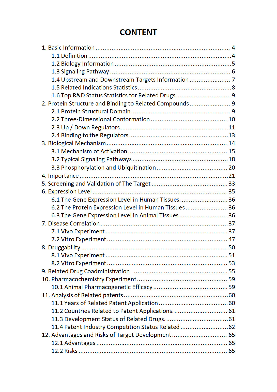
The "TYR Target / Biomarker Review Report" is a customizable review of hundreds up to thousends of related scientific research literature by AI technology, covering specific information about TYR comprehensively, including but not limited to:
• general information;
• protein structure and compound binding;
• protein biological mechanisms;
• its importance;
• the target screening and validation;
• expression level;
• disease relevance;
• drug resistance;
• related combination drugs;
• pharmacochemistry experiments;
• related patent analysis;
• advantages and risks of development, etc.
The report is helpful for project application, drug molecule design, research progress updates, publication of research papers, patent applications, etc. If you are interested to get a full version of this report, please feel free to contact us at BD@silexon.ai
More Common Targets
TYRO3 | TYRO3P | TYROBP | Tyrosine Kinase | Tyrosine-Protein Kinase ABL | Tyrosine-Protein Kinases Src | Tyrosyl-DNA phosphodiesterase TDP | TYRP1 | TYSND1 | TYW1 | TYW1B | TYW3 | U2 small nuclear ribonucleoprotein auxiliary factor | U2AF1 | U2AF1L4 | U2AF2 | U2SURP | U3 small nucleolar ribonucleoprotein (U3 snoRNP) complex | U5 small nuclear ribonucleoprotein complex | U7 snRNP complex | UACA | UAP1 | UAP1L1 | UBA1 | UBA2 | UBA3 | UBA5 | UBA52 | UBA52P1 | UBA6 | UBA6-DT | UBA7 | UBAC1 | UBAC2 | UBAC2-AS1 | UBALD1 | UBALD2 | UBAP1 | UBAP1L | UBAP2 | UBAP2L | UBASH3A | UBASH3B | UBB | UBBP1 | UBBP2 | UBBP4 | UBC | UBD | UBDP1 | UBE2A | UBE2B | UBE2C | UBE2CP3 | UBE2CP4 | UBE2D1 | UBE2D2 | UBE2D3 | UBE2D3P1 | UBE2D4 | UBE2DNL | UBE2E1 | UBE2E2 | UBE2E3 | UBE2F | UBE2F-SCLY | UBE2FP1 | UBE2G1 | UBE2G2 | UBE2H | UBE2HP1 | UBE2I | UBE2J1 | UBE2J2 | UBE2K | UBE2L1 | UBE2L3 | UBE2L6 | UBE2M | UBE2MP1 | UBE2N | UBE2NL | UBE2O | UBE2Q1 | UBE2Q2 | UBE2Q2P1 | UBE2Q2P11 | UBE2Q2P13 | UBE2Q2P16 | UBE2Q2P2 | UBE2QL1 | UBE2R2 | UBE2R2-AS1 | UBE2S | UBE2T | UBE2U | UBE2V1 | UBE2V1P2 | UBE2V1P9 | UBE2V2


