ATP5PD: A Potential Drug Target for Muscle Pain and Muscular Dysfunction


ATP5PD: A Potential Drug Target for Muscle Pain and Muscular Dysfunction
Introduction
ATP (adenylyl cyclic nucleotide) is a crucial energy source for all living organisms, and its production and breakdown are closely regulated in the body. One of the key enzyme involved in ATP synthesis is ATP synthase (ATP synthase complex), which catalyzes the conversion ofADP (adenylyl cyclic dinucleotide) and phosphate to ATP. H+ transporting ATP synthase (H+-ATP synthase) is a subunit of the ATP synthase complex that is responsible for transporting H+ ions from the cytosol to the mitochondrial inner membrane during the ATP synthesis process. The Fo complex, which consists of the E1 subunit (E1), E2 subunit (E2), and E3 subunit (E3), is another subunit of the ATP synthase complex that is involved in ATP synthesis and breakdown. In this article, we will discuss ATP5PD, a potential drug target for muscle pain and muscular dysfunction due to its involvement in the H+-ATP synthase complex and its potential as a therapeutic approach.
The Importance of ATP Synthesis in Muscle Pain and Muscular Dysfunction
Muscle pain and dysfunction are common conditions that affect millions of people worldwide, including those with chronic muscle pain, muscle weakness, and muscle breakdown. These conditions can be caused by a variety of factors, including aging, genetic mutations, and certain medications. One of the underlying mechanisms behind muscle pain and dysfunction is the decreased production of ATP, which can lead to muscle fatigue and decreased muscle contractions.
ATP is a crucial energy source for muscle function, and its production is closely regulated by the H+-ATP synthase complex. The H+-ATP synthase complex is responsible for generating ATP by catalyzing the conversion ofADP and phosphate to ATP, as well as transporting H+ ions from the cytosol to the mitochondrial inner membrane during the ATP synthesis process. The Fo complex, which consists of the E1 subunit (E1), E2 subunit (E2), and E3 subunit (E3), is another subunit of the ATP synthase complex that is involved in ATP synthesis and breakdown.
In muscle cells, ATP production is regulated by various factors, including the levels of calcium ions, national messenger receptor (NMR), and various enzymes. When muscle cells are activated, the H+-ATP synthase complex is activated, and the Fo complex is recruited to the site of the ATP synthesis to assist in the process. However, muscle cells can become dependent on ATP, and a decrease in ATP production can lead to muscle fatigue and decreased muscle contractions.
The Potential Role of ATP5PD in Muscle Pain and Muscular Dysfunction
The H+-ATP synthase complex is a critical enzyme involved in ATP production, and its activity is regulated by various factors, including the levels of calcium ions, national messenger receptor (NMR), and various enzymes. ATP5PD, a potential drug target for muscle pain and muscular dysfunction, is a subunit of the H+-ATP synthase complex that is involved in ATP synthesis and breakdown.
ATP5PD is a 25kDa protein that is composed of three subunits: E1, E2, and E3. It is located in the cytosol and is responsible for participating in the ATP synthesis process by catalyzing the conversion ofADP and phosphate to ATP, as well as transporting H+ ions from the cytosol to the mitochondrial inner membrane during the ATP synthesis process.
Studies have shown that ATP5PD is involved in various cellular processes, including muscle contraction, muscle relaxation, and the regulation of ion channels. It has also been shown to play a role in pain signaling, inflammation, and neurodegeneration. In addition,
Protein Name: ATP Synthase Peripheral Stalk Subunit D
Functions: Mitochondrial membrane ATP synthase (F(1)F(0) ATP synthase or Complex V) produces ATP from ADP in the presence of a proton gradient across the membrane which is generated by electron transport complexes of the respiratory chain. F-type ATPases consist of two structural domains, F(1) - containing the extramembraneous catalytic core, and F(0) - containing the membrane proton channel, linked together by a central stalk and a peripheral stalk. During catalysis, ATP synthesis in the catalytic domain of F(1) is coupled via a rotary mechanism of the central stalk subunits to proton translocation. Part of the complex F(0) domain and the peripheric stalk, which acts as a stator to hold the catalytic alpha(3)beta(3) subcomplex and subunit a/ATP6 static relative to the rotary elements
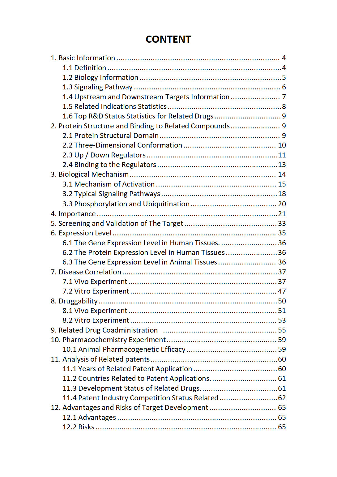
The "ATP5PD Target / Biomarker Review Report" is a customizable review of hundreds up to thousends of related scientific research literature by AI technology, covering specific information about ATP5PD comprehensively, including but not limited to:
• general information;
• protein structure and compound binding;
• protein biological mechanisms;
• its importance;
• the target screening and validation;
• expression level;
• disease relevance;
• drug resistance;
• related combination drugs;
• pharmacochemistry experiments;
• related patent analysis;
• advantages and risks of development, etc.
The report is helpful for project application, drug molecule design, research progress updates, publication of research papers, patent applications, etc. If you are interested to get a full version of this report, please feel free to contact us at BD@silexon.ai
More Common Targets
ATP5PDP3 | ATP5PF | ATP5PO | ATP6 | ATP6AP1 | ATP6AP1-DT | ATP6AP1L | ATP6AP2 | ATP6V0A1 | ATP6V0A2 | ATP6V0A4 | ATP6V0B | ATP6V0C | ATP6V0CP1 | ATP6V0CP3 | ATP6V0D1 | ATP6V0D1-DT | ATP6V0D2 | ATP6V0E1 | ATP6V0E1P1 | ATP6V0E2 | ATP6V0E2-AS1 | ATP6V1A | ATP6V1B1 | ATP6V1B2 | ATP6V1C1 | ATP6V1C2 | ATP6V1D | ATP6V1E1 | ATP6V1E2 | ATP6V1F | ATP6V1FNB | ATP6V1G1 | ATP6V1G1P1 | ATP6V1G2 | ATP6V1G2-DDX39B | ATP6V1G3 | ATP6V1H | ATP7A | ATP7B | ATP8 | ATP8A1 | ATP8A2 | ATP8B1 | ATP8B1-AS1 | ATP8B2 | ATP8B3 | ATP8B4 | ATP8B5P | ATP9A | ATP9B | ATPAF1 | ATPAF2 | ATPase | ATPSCKMT | ATR | ATRAID | Atrial natriuretic peptide (ANP) receptor | ATRIP | ATRN | ATRNL1 | ATRX | ATXN1 | ATXN10 | ATXN1L | ATXN2 | ATXN2L | ATXN3 | ATXN3L | ATXN7 | ATXN7L1 | ATXN7L2 | ATXN7L3 | ATXN7L3B | ATXN8OS | Augmin | AUH | AUNIP | AUP1 | AURKA | AURKAIP1 | AURKAP1 | AURKB | AURKC | Aurora Kinase | AUTS2 | AVEN | AVIL | AVL9 | AVP | AVPI1 | AVPR1A | AVPR1B | AVPR2 | AWAT1 | AWAT2 | AXDND1 | AXIN1 | AXIN2 | AXL


