Drug Target and Biomarker: ATM (G472)


Drug Target and Biomarker: ATM
Low-dose X-ray exposure can lead to cell death, and the ATM molecule plays a role in cellular signal transduction in this process.
ATM activation involves a complex system of biochemical reactions and interactions with other proteins, both in undamaged chromatin and damaged chromatin.
In the life cycle of Epstein-Barr virus (EBV), ATM plays a central role in the replication compartment during the lytic cycle and is regulated by EBV during latency.
ATM is involved in the activation of various proteins and pathways at DNA double-strand break (DSB) sites, including phosphorylation of H2AX and recruitment of MDC1 and 53BP1.
Culturing cells with certain drugs can lead to the development of drug-resistant phenotypes, and the ATM pathway related to DNA repair is found to be increased in cells cultured with tyrosine kinase inhibitors (TKIs) compared to rapalogs.
These viewpoints provide insights into the involvement of ATM in different cellular processes and its relevance in response to external factors such as X-ray exposure or drug treatment.
From the provided information, it can be understood that ATM plays a crucial role in the DNA damage response (DDR) and cellular homeostasis. Normal cells have low expression levels of MELK and effective DNA damage checkpoints, which maintain a balance between DDR and senescence or apoptosis. In cancer cells, overexpression of MELK inhibits ATM-mediated DDR, leading to an accumulation of stalled replication forks, formation of double-strand breaks (DSBs), and genetic instability. Inhibition and degradation of MELK reactivates/enhances the DDR in cancer cells, resulting in growth arrest and senescence.
ATM is involved in repairing double-strand breaks and relies on other proteins such as PARP, BRCA1, and BRCA2 for efficient repair. Inhibition or mutations of these proteins can lead to cell death. Moreover, an approach known as "synthetic lethality" targets specific DDR deficiencies in cancer cells, offering the potential for single-agent activity in treating these cells.
ATM is activated in response to DNA damage, leading to widespread DDR signaling in cellular double-strand breaks. However, in viral DNA, ATM activation is limited to the recruitment of DSB-sensing complexes, without downstream phosphorylation of transducer and effector proteins.
Ubiquitination of ATMIN increases in response to DNA damage, leading to its degradation and release of ATM. Activated ATM phosphorylates various targets to facilitate DNA repair. Loss of function of PPARγ inhibits ATMIN ubiquitination, suppressing ATM activation and signaling, which can result in persistent DNA lesions and genomic instability.
Specific types of DNA damage, such as mismatches, single-strand breaks (SSBs), or double-strand breaks (DSBs), activate different DDR pathways and repair mechanisms. PARP enzymes play a key role in SSB repair, while DSBs are predominantly repaired through non-homologous end joining (NHEJ) or homologous recombination (HR) pathways. ATR, ATM, and DNA-PK are crucial kinases in DDR signaling and maintaining replication fork stability. Drugs targeting these key components of DDR pathways are being tested in clinical trials.
In summary, ATM is a critical player in the DDR, with its activation and phosphorylation of targets facilitating DNA repair and maintaining genomic stability. Overexpression of MELK in cancer cells inhibits ATM-mediated DDR, leading to genetic instability. Inhibition and degradation of MELK can reactivate the DDR, sensitizing cancer cells to DNA damage. Additionally, targeting specific DDR deficiencies or components, such as PARP, BRCA1, BRCA2, etc., can lead to cell death or synthetic lethality in cancer cells. Clinical trials are underway to explore the therapeutic potential of drugs targeting key DDR components.
According to the provided information, both EGF-activated mTOR and hypoxia-induced ATM signaling contribute to the regulation of KPNA2 transcription in lung ADC cells. These signaling pathways suppress IRF1 expression and enhance E2F1 expression, leading to the overexpression of KPNA2. EGF treatment was found to enhance KPNA2 and E2F1 expression, while suppressing IRF1 in A549 ADC cells. However, in NCI-H460 LCC cells, EGF treatment increased IRF1 levels. Additionally, hypoxia induced KPNA2 and E2F1 expression, while reducing IRF1 expression in A549 ADC cells, but not in NCI-H460 LCC cells. Overall, the activation of both mTOR and ATM signaling pathways promotes KPNA2 transcription by regulating the expression levels of IRF1 and E2F1 in lung ADC cells.
Protein Name: ATM Serine/threonine Kinase
Functions: Serine/threonine protein kinase which activates checkpoint signaling upon double strand breaks (DSBs), apoptosis and genotoxic stresses such as ionizing ultraviolet A light (UVA), thereby acting as a DNA damage sensor (PubMed:9733514, PubMed:10550055, PubMed:10839545, PubMed:10910365, PubMed:12556884, PubMed:14871926, PubMed:15456891, PubMed:15448695, PubMed:15916964, PubMed:17923702). Recognizes the substrate consensus sequence [ST]-Q (PubMed:9733514, PubMed:10550055, PubMed:10839545, PubMed:10910365, PubMed:12556884, PubMed:14871926, PubMed:15456891, PubMed:15448695, PubMed:15916964, PubMed:17923702). Phosphorylates 'Ser-139' of histone variant H2AX at double strand breaks (DSBs), thereby regulating DNA damage response mechanism (By similarity). Also plays a role in pre-B cell allelic exclusion, a process leading to expression of a single immunoglobulin heavy chain allele to enforce clonality and monospecific recognition by the B-cell antigen receptor (BCR) expressed on individual B-lymphocytes. After the introduction of DNA breaks by the RAG complex on one immunoglobulin allele, acts by mediating a repositioning of the second allele to pericentromeric heterochromatin, preventing accessibility to the RAG complex and recombination of the second allele. Also involved in signal transduction and cell cycle control. May function as a tumor suppressor. Necessary for activation of ABL1 and SAPK. Phosphorylates DYRK2, CHEK2, p53/TP53, FBXW7, FANCD2, NFKBIA, BRCA1, CTIP, nibrin (NBN), TERF1, UFL1, RAD9, UBQLN4 and DCLRE1C (PubMed:9843217, PubMed:9733515, PubMed:10550055, PubMed:10766245, PubMed:10839545, PubMed:10910365, PubMed:10802669, PubMed:10973490, PubMed:11375976, PubMed:12086603, PubMed:15456891, PubMed:19965871, PubMed:30612738, PubMed:30886146, PubMed:26774286). May play a role in vesicle and/or protein transport. Could play a role in T-cell development, gonad and neurological function. Plays a role in replication-dependent histone mRNA degradation. Binds DNA ends. Phosphorylation of DYRK2 in nucleus in response to genotoxic stress prevents its MDM2-mediated ubiquitination and subsequent proteasome degradation (PubMed:19965871). Phosphorylates ATF2 which stimulates its function in DNA damage response (PubMed:15916964). Phosphorylates ERCC6 which is essential for its chromatin remodeling activity at DNA double-strand breaks (PubMed:29203878). Phosphorylates TTC5/STRAP at 'Ser-203' in the cytoplasm in response to DNA damage, which promotes TTC5/STRAP nuclear localization (PubMed:15448695). Also involved in pexophagy by mediating phosphorylation of PEX5: translocated to peroxisomes in response to reactive oxygen species (ROS), and catalyzes phosphorylation of PEX5, promoting PEX5 ubiquitination and induction of pexophagy (PubMed:26344566)
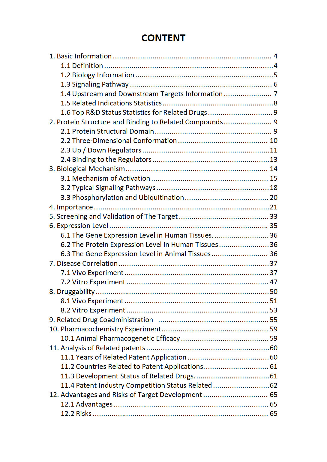
The "ATM Target / Biomarker Review Report" is a customizable review of hundreds up to thousends of related scientific research literature by AI technology, covering specific information about ATM comprehensively, including but not limited to:
• general information;
• protein structure and compound binding;
• protein biological mechanisms;
• its importance;
• the target screening and validation;
• expression level;
• disease relevance;
• drug resistance;
• related combination drugs;
• pharmacochemistry experiments;
• related patent analysis;
• advantages and risks of development, etc.
The report is helpful for project application, drug molecule design, research progress updates, publication of research papers, patent applications, etc. If you are interested to get a full version of this report, please feel free to contact us at BD@silexon.ai
More Common Targets
ATMIN | ATN1 | ATOH1 | ATOH7 | ATOH8 | ATOSA | ATOSB | ATOX1 | ATOX1-AS1 | ATP Synthase, H+ Transporting, Mitochondrial F0 complex | ATP synthase, H+ transporting, mitochondrial F1 complex | ATP-Binding Cassette (ABC) Transporter | ATP-dependent 6-phosphofructokinase | ATP10A | ATP10B | ATP10D | ATP11A | ATP11A-AS1 | ATP11AUN | ATP11B | ATP11C | ATP12A | ATP13A1 | ATP13A2 | ATP13A3 | ATP13A3-DT | ATP13A4 | ATP13A5 | ATP13A5-AS1 | ATP1A1 | ATP1A1-AS1 | ATP1A2 | ATP1A3 | ATP1A4 | ATP1B1 | ATP1B2 | ATP1B3 | ATP1B4 | ATP23 | ATP2A1 | ATP2A1-AS1 | ATP2A2 | ATP2A3 | ATP2B1 | ATP2B1-AS1 | ATP2B2 | ATP2B3 | ATP2B4 | ATP2C1 | ATP2C2 | ATP4A | ATP4B | ATP5F1A | ATP5F1B | ATP5F1C | ATP5F1D | ATP5F1E | ATP5F1EP2 | ATP5IF1 | ATP5MC1 | ATP5MC1P3 | ATP5MC2 | ATP5MC3 | ATP5ME | ATP5MF | ATP5MG | ATP5MGL | ATP5MJ | ATP5MK | ATP5PB | ATP5PBP5 | ATP5PD | ATP5PDP3 | ATP5PF | ATP5PO | ATP6 | ATP6AP1 | ATP6AP1-DT | ATP6AP1L | ATP6AP2 | ATP6V0A1 | ATP6V0A2 | ATP6V0A4 | ATP6V0B | ATP6V0C | ATP6V0CP1 | ATP6V0CP3 | ATP6V0D1 | ATP6V0D1-DT | ATP6V0D2 | ATP6V0E1 | ATP6V0E1P1 | ATP6V0E2 | ATP6V0E2-AS1 | ATP6V1A | ATP6V1B1 | ATP6V1B2 | ATP6V1C1 | ATP6V1C2 | ATP6V1D


