CAB39L: A Calcium Binding Protein with Potential as a Drug Target or Biomarker


CAB39L: A Calcium Binding Protein with Potential as a Drug Target or Biomarker
Introduction
Calcium homeostasis is a critical regulatory process in various physiological processes, including muscle contractions, nerve function, and brain development. The regulation of calcium homeostasis is achieved by a complex network of proteins, including calcium-binding proteins (CABs), which play a vital role in the bioavailability of cisplatin and its role in chemotherapy drugs. CABs are a family of transmembrane proteins that can interact with calcium ions and regulate various cellular processes. In this article, we will focus on CAB39L, a calcium-binding protein that has potential as a drug target or biomarker.
CAB39L: Structure and Function
CAB39L is a 29 kDa protein that belongs to the CAB family. It is expressed in various tissues, including brain, heart, and muscle. CAB39L functions as a calcium-binding protein by interacting with calcium ions in the cytosol. This interaction is essential for the regulation of various cellular processes, including muscle contractions, nerve function, and brain development.
CAB39L is composed of a variable region (V1-V3) and an N-terminal region (N). The V1 region contains a unique N-terminal domain, known as the CAB39L-specific domain, which is responsible for the protein's calcium-binding affinity. The CAB39L-specific domain is a short, N-terminal alpha-helical domain that is rich in conserved electrolytic features, including a positively charged amino acid residue (D) and a negatively charged amino acid residue (E) at its N -terminus.
The N-terminal region (N) of CAB39L contains a long amino acid sequence that is involved in the formation of a disulfide bond. This region also contains a single transmembrane segment (TMS) that is responsible for the protein's ability to interact with calcium ions. The TMS is composed of a long alpha-helical segment (alpha-helices) and a short beta-sheet segment (beta-sheet) that are connected by a disulfide bond.
CAB39L Interactions with Calcium Ions
CAB39L is primarily regulated by the cytosol calcium ion (Ca2+) concentration. The cytosol is the space between the mitochondria and the endoplasmic reticulum, and it is a critical environment for the regulation of various cellular processes. The cytosol is the main site of Ca2+ storage and utilization, and it is where the majority of Ca2+ ions are generated through the uptake of dietary calcium.
CAB39L is a calcium-binding protein that can interact with Ca2+ ions in the cytosol. The interaction between CAB39L and Ca2+ ions is critical for the regulation of various cellular processes, including muscle contractions, nerve function, and brain development.
CAB39L has been shown to play a role in the regulation of muscle contractions. In myotonic dystrophy, a genetic disorder characterized by muscle weakness and degenerative muscle wasting, CAB39L has been shown to be involved in the regulation of muscle contractions. Studies have shown that individuals with myotonic dystrophy have lower levels of CAB39L in their muscle tissue compared to healthy individuals, and that these individuals also have reduced muscle function.
CAB39L has also been shown to play a role in the regulation of nerve function. In neurodegenerative diseases, such as Alzheimer's disease, CAB39L has been shown to be involved in the regulation of nerve function. Studies have shown that individuals with neurodegenerative diseases have lower levels of CAB39L in their brain tissue compared to healthy individuals, and that these individuals also have reduced nerve function.
CAB39L has also been shown to play a role in the regulation of brain development. During brain development, CAB39L has been shown to be involved in the regulation of neurotransmitter release from neurons. Studies have shown that individuals with brain development
Protein Name: Calcium Binding Protein 39 Like
Functions: Component of a complex that binds and activates STK11/LKB1. In the complex, required to stabilize the interaction between CAB39/MO25 (CAB39/MO25alpha or CAB39L/MO25beta) and STK11/LKB1 (By similarity)
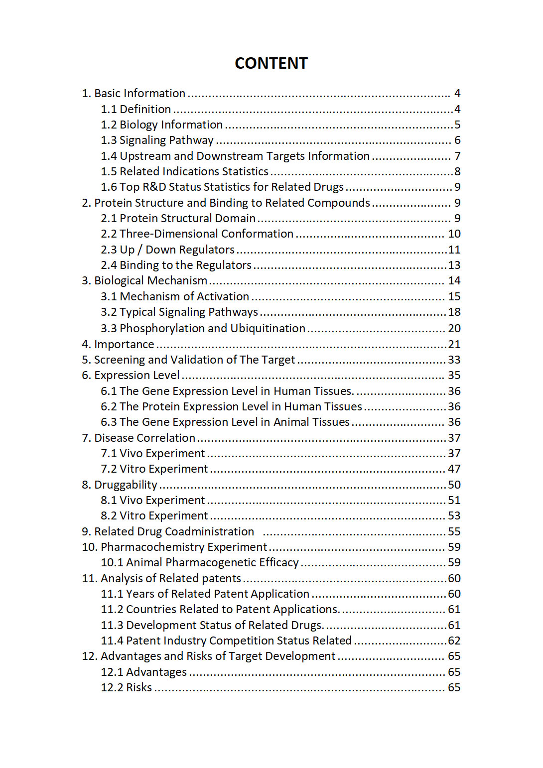
The "CAB39L Target / Biomarker Review Report" is a customizable review of hundreds up to thousends of related scientific research literature by AI technology, covering specific information about CAB39L comprehensively, including but not limited to:
• general information;
• protein structure and compound binding;
• protein biological mechanisms;
• its importance;
• the target screening and validation;
• expression level;
• disease relevance;
• drug resistance;
• related combination drugs;
• pharmacochemistry experiments;
• related patent analysis;
• advantages and risks of development, etc.
The report is helpful for project application, drug molecule design, research progress updates, publication of research papers, patent applications, etc. If you are interested to get a full version of this report, please feel free to contact us at BD@silexon.ai
More Common Targets
CABCOCO1 | CABIN1 | CABLES1 | CABLES2 | CABP1 | CABP2 | CABP4 | CABP5 | CABP7 | CABS1 | CABYR | CACFD1 | CACHD1 | CACNA1A | CACNA1B | CACNA1C | CACNA1C-AS4 | CACNA1C-IT2 | CACNA1C-IT3 | CACNA1D | CACNA1E | CACNA1F | CACNA1G | CACNA1G-AS1 | CACNA1H | CACNA1I | CACNA1S | CACNA2D1 | CACNA2D1-AS1 | CACNA2D2 | CACNA2D3 | CACNA2D4 | CACNB1 | CACNB2 | CACNB3 | CACNB4 | CACNG1 | CACNG2 | CACNG2-DT | CACNG3 | CACNG4 | CACNG5 | CACNG6 | CACNG7 | CACNG8 | CACTIN | CACTIN-AS1 | CACUL1 | CACYBP | CAD | CADM1 | CADM2 | CADM3 | CADM3-AS1 | CADM4 | CADPS | CADPS2 | CAGE1 | CAHM | CALB1 | CALB2 | CALCA | CALCB | Calcium channel | Calcium release-activated channel (CRAC) | Calcium-activated chloride channel regulators | Calcium-Activated K(Ca) Potassium Channel | CALCOCO1 | CALCOCO2 | CALCR | CALCRL | CALCRL-AS1 | CALD1 | CALHM1 | CALHM2 | CALHM3 | CALHM4 | CALHM5 | CALHM6 | CALM1 | CALM2 | CALM2P1 | CALM2P2 | CALM3 | CALML3 | CALML3-AS1 | CALML4 | CALML5 | CALML6 | Calmodulin | CALN1 | Calpain | Calpain-13 | Calprotectin | CALR | CALR3 | CALU | CALY | CAMK1 | CAMK1D


